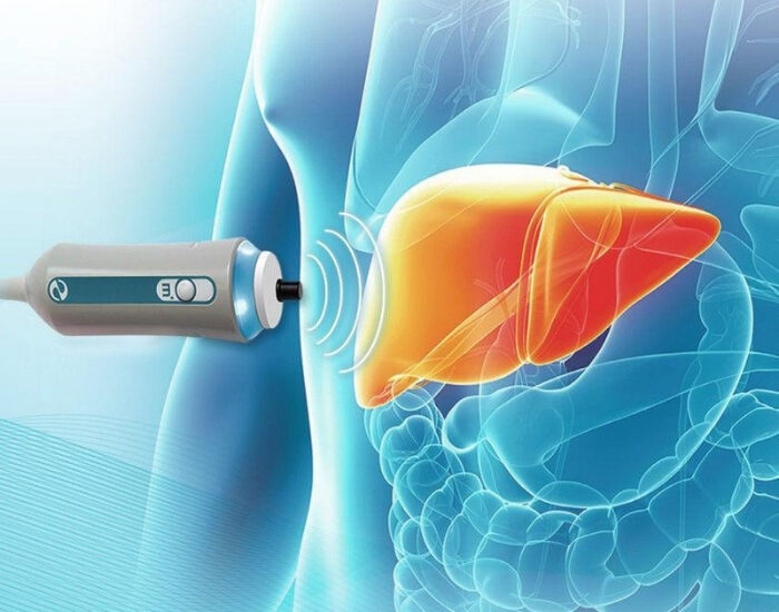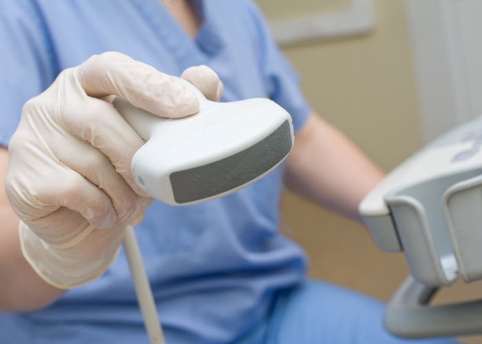
“Diffuse hepatic steatosis” is one of the most common phrases on abdominal ultrasound and CT reports in the United States. It often shows up when the scan was ordered for something else — gallbladder pain, kidney stones, abdominal pain after a trip to the ER.
The phrase sounds alarming, but on its own it describes a finding, not a disease stage. This guide translates the radiology wording, explains what the grades mean and walks through the follow-up a liver specialist would recommend.
Key takeaways
- Diffuse means the fat is spread through the whole liver; hepatic steatosis means fatty liver.
- Mild, moderate and severe describe visible fat, not liver damage.
- Ultrasound cannot reliably show inflammation or scarring.
- A FIB-4 score and FibroScan are the usual next steps.
What Does Diffuse Hepatic Steatosis Mean?
Diffuse hepatic steatosis means fat has built up evenly throughout the liver tissue. “Hepatic” refers to the liver, “steatosis” means abnormal fat storage, and “diffuse” means the change involves the whole organ rather than one area. Medically, steatosis is significant once fat makes up more than about 5% of the liver.
Radiologists may use several phrases for the same finding:
- Diffuse hepatic steatosis
- Diffuse fatty infiltration of the liver
- Increased hepatic echogenicity, consistent with steatosis
- Fatty liver
- Hepatic steatosis with or without hepatomegaly (an enlarged liver)
All of these point to the same basic question your doctor needs to answer: what is causing the fat, and has it affected the liver’s health? For the bigger picture, see what fatty liver disease is.
What Is the Difference Between Diffuse and Focal Steatosis?
Diffuse steatosis affects the whole liver evenly. Focal steatosis is fat in one patch, and focal fatty sparing is a patch of normal liver inside an otherwise fatty liver. Both focal patterns can look like a mass on imaging, so they sometimes need a second test to confirm they are harmless.
- Diffuse steatosis: uniform brightness on ultrasound or uniform low density on CT across the liver.
- Heterogeneous or geographic steatosis: uneven distribution with irregular areas.
- Focal fatty infiltration: a single area of fat, often near the gallbladder or the liver’s blood vessels.
- Focal fatty sparing: a darker “spot” of normal liver surrounded by fat; sometimes reported as a possible lesion.
If your report mentions a focal area or a “lesion,” ask whether an MRI is recommended to be sure it is fat and not something else.
How Do Ultrasound, CT and MRI Show Hepatic Steatosis?
On ultrasound, a fatty liver looks brighter, or more echogenic, than the kidney, and the deeper liver may be harder to see. On a non-contrast CT it looks darker, with low density measured in Hounsfield units. MRI with proton density fat fraction measures liver fat most precisely. None of these scans reliably shows scarring, which needs separate testing.
| Imaging test | What fatty liver looks like | Strengths | Limits |
|---|---|---|---|
| Ultrasound | Liver brighter than the kidney; vessels and diaphragm harder to see | Widely available, no radiation, low cost | Misses small amounts of fat; less accurate with obesity; cannot measure scarring |
| CT scan | Liver darker than the spleen; low attenuation (under about 40 HU on non-contrast CT is specific for steatosis) | Often already done for another reason | Radiation; best on non-contrast scans; not sensitive for mild fat |
| MRI (PDFF) | Fat fraction reported as a percentage; 5% or more indicates steatosis | Most accurate measure of liver fat | Cost and availability; not needed for most patients |
| FibroScan (CAP) | Controlled attenuation parameter score for fat, plus liver stiffness | Measures fat and scarring in one quick office test | Less reliable with very high body weight or fluid in the abdomen |
What Do Mild, Moderate and Severe Diffuse Hepatic Steatosis Mean?
On ultrasound, radiologists commonly grade steatosis by how much the extra brightness hides the structures around and inside the liver. Mild means the liver is brighter but vessels and the diaphragm are still clear; moderate means vessel walls are blurred; severe means the diaphragm and deep liver are hard to see.
Common ultrasound grades of diffuse hepatic steatosis
- Mild (grade 1)Liver brighter than normal; portal vein walls and diaphragm still well seen.
- Moderate (grade 2)Brightness blurs the walls of the portal veins; diaphragm partly obscured.
- Severe (grade 3)Diaphragm and deeper liver poorly seen because sound waves are absorbed.
Does a higher grade mean worse liver disease?
Not necessarily. Grade reflects the amount of fat the ultrasound can see. It does not show inflammation or fibrosis, which are what drive long-term liver risk. In our hepatology clinic in Katy and Cypress, we regularly see patients with “mild” steatosis who have meaningful scarring, and others with “severe” steatosis whose liver stiffness is normal. That is why grading is a starting point, not the answer.
Can ultrasound show scarring or cirrhosis?
Ultrasound can suggest advanced cirrhosis — a nodular surface, an enlarged spleen or fluid in the abdomen — but it misses earlier fibrosis. A “coarse echotexture” on the report may raise the question of fibrosis but does not confirm it. A FibroScan or blood-based score is needed.
Have a report that says hepatic steatosis?
Bring it to Dr. Muhammad Ali Khan, a board-certified gastroenterologist and hepatologist. We can review your imaging and labs and check liver stiffness with FibroScan at our Katy and Cypress offices.
What Causes Diffuse Hepatic Steatosis?
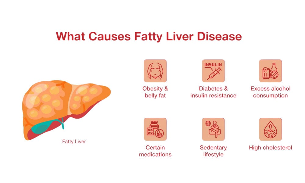
The most common cause is MASLD, fatty liver linked to excess weight, insulin resistance, type 2 diabetes, high triglycerides or high blood pressure. Alcohol use is the next most common cause. Less often, steatosis comes from medicines such as steroids or methotrexate, hepatitis C, rapid weight loss, malnutrition or rare genetic conditions, so your doctor reviews all of these.
- Metabolic (MASLD): the leading cause in Texas and across the U.S.
- Alcohol: alone, or combined with metabolic risk (MetALD).
- Medicines: corticosteroids, methotrexate, amiodarone, tamoxifen, some HIV and psychiatric medicines.
- Infections: especially hepatitis C (genotype 3).
- Nutritional: rapid weight loss, very low protein intake, prolonged IV nutrition.
- Other: underactive thyroid, polycystic ovary syndrome, sleep apnea and rare inherited conditions.
What Should You Do After a Diffuse Hepatic Steatosis Finding?
After a diffuse hepatic steatosis finding, ask your doctor for liver blood tests and a FIB-4 fibrosis score, and review your alcohol use and medicines. Check blood sugar, cholesterol and blood pressure. If the FIB-4 score is not low, arrange a FibroScan or other stiffness test. Seeing a liver specialist is wise if scarring is suspected or enzymes stay raised.
- Liver panel and platelets. ALT, AST and platelets feed into the FIB-4 score. Normal enzymes do not rule out scarring.
- FIB-4 score. Low scores make advanced fibrosis unlikely; indeterminate or high scores call for elastography.
- Screen for other causes. Hepatitis B and C, iron studies and, when indicated, autoimmune or genetic liver tests.
- Metabolic check. A1c or fasting glucose, lipids, blood pressure and waist size.
- FibroScan. Measures stiffness and fat in about 10 minutes. See our FibroScan page.
- MRI or biopsy only when results disagree or another diagnosis is suspected.
If your enzymes are abnormal, our guide to abnormal ALT and AST explains what the numbers can mean.
Can Diffuse Hepatic Steatosis Be Reversed?
Yes, diffuse hepatic steatosis can often be reversed because liver fat responds quickly to change. Losing about 3% to 5% of body weight can reduce steatosis, and losing 7% to 10% or more can improve inflammation and sometimes scarring. Regular exercise, less sugar and alcohol, and good control of diabetes and cholesterol all help the liver recover.
- Aim for steady weight loss of 1–2 pounds a week rather than crash dieting
- Cut sugary drinks, sweets and refined carbohydrates
- Follow a Mediterranean-style eating pattern
- Get about 150 minutes of moderate activity a week
- Limit alcohol, or avoid it if there is any scarring
- Treat diabetes, cholesterol, blood pressure and sleep apnea
For people with MASH and significant fibrosis, prescription treatments are now available, and some diabetes and weight-loss medicines also benefit the liver. A specialist can tell you whether you are a candidate. More detail is in Can fatty liver be reversed?
When Should You See a Liver Specialist for Hepatic Steatosis?
See a gastroenterologist or hepatologist if your FIB-4 or FibroScan suggests scarring, your liver enzymes stay elevated, you have type 2 diabetes with fatty liver, the report mentions a focal lesion or coarse liver, or you have symptoms such as jaundice, belly swelling or easy bruising. Patients from Katy, Cypress, Fulshear, Richmond and the wider Houston area can bring their imaging report and recent labs to the first visit.
Learn more on our fatty liver specialist page and liver specialist page.
Frequently Asked Questions About Diffuse Hepatic Steatosis
Is diffuse hepatic steatosis serious?
By itself, diffuse hepatic steatosis is a common and often reversible finding. It becomes serious if the fat has caused inflammation and scarring.
What is the difference between hepatic steatosis and fatty liver?
They mean the same thing. Hepatic steatosis is the medical term for fat stored in the liver, and fatty liver is the everyday term.
What does mild diffuse hepatic steatosis mean?
Mild means the liver looks somewhat brighter than normal on ultrasound but surrounding structures are still clearly seen. It reflects a smaller amount of visible fat, but it does not rule out scarring, so follow-up testing is still recommended.
Where can I see a liver specialist for hepatic steatosis in Katy or Cypress, TX?
Dr. Muhammad Ali Khan, a board-certified gastroenterologist and hepatologist at United Gastroenterology Associates, evaluates fatty liver and offers FibroScan testing at offices in Katy and Cypress. Call (346) 340-4414.
What sources support this article?
- AASLD — Practice guidance on the clinical assessment and management of NAFLD (2023)
- AASLD — Changes in fatty liver nomenclature (MASLD)
- Radiopaedia — Hepatic steatosis and grading of diffuse hepatic steatosis
- NIDDK — Nonalcoholic fatty liver disease and NASH
Where can you see a gastroenterologist in Katy or Cypress?
United Gastroenterology Associates has two offices: Katy, at 23920 Katy Freeway, Suite 555, next to Memorial Hermann Katy Hospital, and Cypress, at 21216 Northwest Freeway, Suite 640, on US-290. Both are open Monday through Friday, 8:30 AM to 5:00 PM, closed for lunch from 12:00 to 1:30 PM, and serve patients from Fulshear, Richmond and across Greater Houston. Call (346) 340-4414 for either office.
Where is the Katy office?
23920 Katy Freeway, Suite 555
Katy, TX 77494
Memorial Hermann Medical Plaza 1
Where is the Cypress office?
21216 Northwest Freeway, Suite 640
Cypress, TX 77429
On US-290
What should you do next?
If your ultrasound, CT or MRI report mentions diffuse hepatic steatosis, the next step is finding out why it is there and whether the liver has any scarring. Dr. Muhammad Ali Khan, a board-certified gastroenterologist and hepatologist, sees adults from Katy, Cypress, Fulshear, Richmond and across Greater Houston. Call (346) 340-4414 or request an appointment today.